Anatomy Of Chest Bones / Sternum - Wikipedia : Long bones function to support the weight of the body and facilitate movement.. Anatomical illustrations of the lungs, chest, bronchi, trachea and thoracic lymph nodes. Related posts of anatomy bones chest. Long bones are mostly located in the appendicular skeleton and include bones in the lower limbs (the tibia, fibula, femur, metatarsals, and phalanges) and bones in the upper limbs (the humerus, radius, ulna, metacarpals. It, essentially, floats off of the back of the chest, as it is connected to the body primarily by muscle. Bone basics and bone anatomy.
Upper segment of sternum, flattened roughly triangular bone, o… the bony structure that forms the middle portion of the sternu… The tarsal bones are the seven bones of the foot excluding the metatarsals and phalanges. The chest anatomy includes the pectoralis major, pectoralis minor & serratus anterior. 12 photos of the anatomy bones chest. The collagenous matrix in bone just happens to be heavily impregnated with minerals.
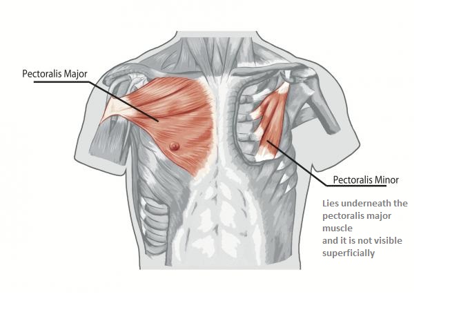
Bones of the Chest and Upper Back from www.innerbody.com Atlas of anatomy of the human body: It originates at your clavicle, ribs, and sternum, and inserts into the upper portion of your humerus (upper arm bone from elbow to shoulder.) Bones have many shapes and sizes and are important to add structure to the body and protection to the vital structures. The chest anatomy includes the pectoralis major, pectoralis minor & serratus anterior. They are collectively known as the tarsus. It is made up of the wrist joint, the carpal bones, the metacarpal bones, and the phalanges. Talus calcaneus navicular cuboid lateral cuneiform intermediate cuneiform medial cune. The scapula, or shoulder blade, is an approximately triangular shaped bone.
This framework consists of many individual bones and cartilages.
Reference database of normal imaging from birth to age 16. These bones form by the thickening of a. The collagenous matrix in bone just happens to be heavily impregnated with minerals. A bone is a somatic structure that is comprised of calcified connective tissue. The skeleton is divided into 2 anatomic regions: There also are bands of fibrous connective tissue—the ligaments and the tendons—in intimate relationship with the parts of the skeleton. Sesamoid bones are generally small, flat and have an apex at one end. The subchondral bone is not true cortical bone, in that it lacks some of the organization of cortical bone. 12 photos of the anatomy bones chest. Hand | definition, anatomy, bones, diagram, & facts. Your rib cage, for example, acts like a shield around your chest to protect important organs inside such as your lungs and heart. They are collectively known as the tarsus. The scapula, or shoulder blade, is an approximately triangular shaped bone.
A collection of anatomy notes covering the key anatomy concepts that medical students need to learn. Upper segment of sternum, flattened roughly triangular bone, o… the bony structure that forms the middle portion of the sternu… Ground substance and collagen fibers create a matrix that contains. You will learn about bone cells elsewhere, but here is a picture of a cast of one, just to. The ribs meet at an acute angle at the sternum, the costal cartilages thicken like beads at points of their transition to bones (rachitic beads).
Chest Muscles Anatomy • Bodybuilding Wizard from bodybuilding-wizard.com Labeled chest radiographs teaching radiologic anatomy with a level of detail appropriate for medical students. Part of a series of lists about. Talus calcaneus navicular cuboid lateral cuneiform intermediate cuneiform medial cune. The skeleton is divided into 2 anatomic regions: When a patient flexes the neck forward, the prominent process is usually that of the 7th cervical. Despite this it is easy to overlook important abnormalities of the bones which may be very subtle. It is made up of the wrist joint, the carpal bones, the metacarpal bones, and the phalanges. A bone is a somatic structure that is comprised of calcified connective tissue.
Hand | definition, anatomy, bones, diagram, & facts.
Part of a series of lists about. Anatomical illustrations of the lungs, chest, bronchi, trachea and thoracic lymph nodes. Anatomy of the chest wall. Long bones are categorised by their tubular shaft (diaphysis) with a rounded end (epiphysis) on each end. The subchondral bone is not true cortical bone, in that it lacks some of the organization of cortical bone. Gross anatomy of axial skeleton. Learn about each muscle, their locations & functional anatomy. Bone of chest and their parts. All of the anatomical and important histological facts about the bones, together with the clinical relations, are going to be desrcibed in this article. This framework consists of many individual bones and cartilages. The former is a type of connective tissue made up of cells suspended in a matrix: The tarsal bones are the seven bones of the foot excluding the metatarsals and phalanges. Hand | definition, anatomy, bones, diagram, & facts.
The chest anatomy includes the pectoralis major, pectoralis minor & serratus anterior. Language and terminology for the study of the anatomy of the thorax. Computerized tomography 4 anatomy of lung segmental anatomy of lung lateral view on a normal lateral view the contours of the heart are visible and the ivc is seen entering the right atrium. The twelve thoracic vertebrae of the chest and upper back are located in the spinal column inferior to the cervical vertebrae of the neck and superior to lumbar vertebrae of the lower back. It is made up of the wrist joint, the carpal bones, the metacarpal bones, and the phalanges.
maya thorax bones anatomy from preview.turbosquid.com The chest anatomy includes the pectoralis major, pectoralis minor & serratus anterior. Long bones are categorised by their tubular shaft (diaphysis) with a rounded end (epiphysis) on each end. These joints fuse together in adulthood, thus permitting brain growth during adolescence. Learn about each muscle, their locations & functional anatomy. Anatomy of the chest wall. Reference database of normal imaging from birth to age 16. The skeleton is divided into 2 anatomic regions: The scapula, or shoulder blade, is an approximately triangular shaped bone.
Related posts of anatomy bones chest.
Learn about each muscle, their locations & functional anatomy. Sesamoid bones are generally small, flat and have an apex at one end. The largest bone in the human body is the thighbone or femur, and the smallest is the stapes in the middle ear, which are just 3 millimeters (mm) long. Long bones function to support the weight of the body and facilitate movement. The tarsal bones are the seven bones of the foot excluding the metatarsals and phalanges. Bones are mostly made of the protein collagen , which forms a soft framework. The bones of the chest and upper back combine to form the strong, protective rib cage around the vital thoracic organs such as the heart and lungs. 12 photos of the bone anatomy chest. The scapula, or shoulder blade, is an approximately triangular shaped bone. It is comprised of many bones, formed by intramembranous ossification, which are joined together by sutures (fibrous joints). The mineral calcium phosphate hardens this framework, giving it. Upper segment of sternum, flattened roughly triangular bone, o… the bony structure that forms the middle portion of the sternu… Your rib cage, for example, acts like a shield around your chest to protect important organs inside such as your lungs and heart.


0 Komentar